Point of Care Ultrasound (POCUS)Training with MedicScan
For African Medical Professionals
Obstetrics and Gynaecology POCUS Training
Feature in MedicScan
.jpg?width=4032&height=3024&name=iOS%20%E3%81%AE%E7%94%BB%E5%83%8F%20(10).jpg)
CPD
Accreditation
POCUS training is CPD accredited in Kenya. Healthcare Professionals participating in the training will receive CPD points from their respective councils.
Soon, the CPD Program will be available to participants in other countries.

Multi Country
We are expanding our services to various African countries, mainly in East Africa, and throughout the entire continent. In fact, doctors from Tanzania and Zambia have taken this training.

Access to Anytime for your skills
We are creating an environment for healthcare professionals to have access to equipment and training at any time.
Course Milestones
Price Option:
1. Training only: 25,000KES
2. Training and POCUS Text Book: 38,000KES
3. Training, POCUS Text Book and Flash Drive: 39,000KES
Mode: Online and Physical hands on Physical training Centre: B7C, Chelezo Apartments, Kindaruma Rd, NRB.
Contact us at +254 726 997 407 or medicscan@aa-healthdynamics.com
This 3 months skillset Point of Care Ultrasound (POCUS) training shall be delivered online by Certified Medical Trainers who graduated POCUS Training skill set Program Designed by Dr.Taro Minami, MD, FACP, FCCP, Director of Medical Simulation and Point-of-Care Ultrasound Training, Department of Medicine, Kent Hospital and Care New England, Associate Professor of Medicine, Clinician Educator, Associate Professor of Medical Science, Clinician Educator.The Warren Alpert Medical School of Brown University, and hands on training taught physically by Kenyan POCUS trainers.
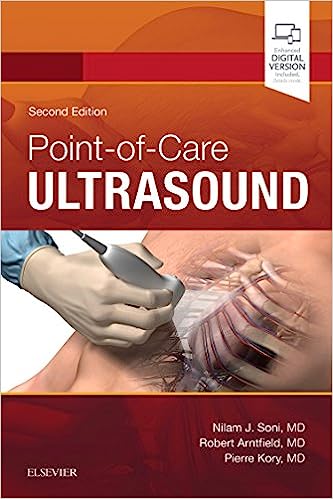
Through lectures, videos and hands on sessions the course aims to offer you deeper insight in application of point of care Ultrasound skills in your practice.
Point of Care Ultrasound (POCUS) will be certified for certificate and CPD for KMPDC and PPB members.

(1) Complete Online Learning Modules
(a) Principles, Knobology, Artifacts
(i) Trainees will learn basic principles of ultrasound, artifact, and knobology via online learning module.
(ii) After learning the online module, they will check their understanding by Quizzes (post-test).
(b) Focused Cardiac Ultrasound (FoCUS)
(i) Trainees will learn focused cardiac ultrasound via online learning module.
(ii) After learning the on-line module, they will check their understanding by Quizzes (post-test).
(c) Lung and Thoracic Ultrasound
(i) Trainees will learn basic principles of lung and thoracic ultrasound, particularly with BLUE-protocol via online learning module.
(ii) After learning the online module, they will check their understanding by Quizzes (post-test).
(d) Abdominal Ultrasound
(i) Trainees will learn essential abdominal ultrasound, particularly with the concept of FAST examination via online learning module.
(ii) After learning the online module, they will check their understanding by Quizzes (post-test).
(e) Vascular Ultrasound
(i) Trainees will learn vascular diagnostic ultrasound, mainly assessment of venous thromboembolic disease.
(ii) After learning the online module, they will check their understanding by Quizzes (post-test).
(f) Assessment of Shock
(i) Trainees will learn assessment of shock by point-of-care ultrasound. This should be learnt upon understanding (a) – (e) as they learn how we incorporate image acquisition and interpretation into clinical integration.

(2) Complete Hands-on Training (5 sessions)
(a) Cardiac Examination
(i) Trainees will learn 5 basic views (parasternal long and short axis view, apical four chamber view, subcostal four chamber and IVC view) via hands-on training supervised by myself and/or by other instructor(s)
(b) Lung Examination
(i) Trainees will learn 3 images: lung sliding with A-lines, Right hemidiaphragm, Left hemidiaphragm) via hands-on training supervised by myself and/or by other instructor(s)
(c) Abdominal Examination
(i) Trainees will learn 4 images
(1) RUQ: Liver, hepatorenal recess, Right kidney,
(2) LUQ, Spleen, splenorenal recess, Left Kidney,
(3) Abdominal Aorta (longitudinal) view,
(4) Bladder – Transverse via hands-on training supervised by myself and/or by other instructor(s).
(d) Vascular Examination
(i) Trainees will learn vascular diagnostics by hands-on training – femoral vein and internal jugular vein – supervised by Dr. Taro Minami and/or by other instructor(s)

(3) Complete Image Portfolio:
14 images for one case, total of 2 cases (total 14 images x 2 = 28 images)
Trainees will obtain images as delineated below from patients they examine. Each image will be de-identified and then sent to me for further evaluation – each image will be assessed with comments – and if not meeting criteria, they are asked to redo the images as needed.
(a) Cardiac
5 images: Parasternal long axis, Parasternal short axis, Apical 4 chamber view, Subcostal 4 chamber view, Subcostal IVC view
(b) Lung
3 images: lung sliding with A-lines, Right hemidiaphragm, Left hemidiaphragm
(c) Abdomen
4 images: RUQ: Liver, hepatorenal recess, Right kidney, LUQ, Spleen, splenorenal recess, Left Kidney, Abdominal Aorta (longitudinal) view, Bladder - Transverse
(d) Vascular
2 images: Common Femoral Vein with compression, Popliteal vein with compression
Once trainee fulfills all the criteria (1) - (3), then can proceed to the examinations (4) and (5):

(4) Final Examinations:
Knowledge and Image interpretation examinations are performed online with multiple-choice format. Details will be communicated to the class. To pass each examination, trainees need to answer at least 80% correctly. Upon completion of knowledge and image interpretation examinations, trainees proceed to the final skill examination, which will be test their skills by observing their scanning of a model.


%20(1).png)